Select the option that best describes what you are looking for
Boost oncology drug discovery with XenoBase®, featuring the largest cell line selection and exclusive 3D organoid models. Benefit from OrganoidXplore™ and OmniScreen™ for rapid, in-depth analysis.
Enhance drug development with our validated in vivo models, in vitro/ex vivo assays, and in silico modeling. Tailored solutions to optimize your candidates.
Experience ISO-certified biobanking quality. Access top biospecimens from a global clinical network, annotated by experts for precise research.
Leverage our global labs and 150+ scientists for fast, tailored project execution. Benefit from our expertise, cutting-edge tech, and validated workflows for reliable data outcomes.
Harness your data and discover biomarkers with our top bioinformatics expertise. Maximize data value and gain critical insights to accelerate drug discovery and elevate projects.
Accelerate innovative cancer treatments with our advanced models and precise drug screening for KRAS mutations, efficiently turning insights into clinical breakthroughs.
Advance translational pharmacology with our diverse pre-clinical models, robust assays, and data science-driven biomarker analysis, multi-omics, and spatial biology.
Our suite integrates preclinical solutions, bioanalytical read-outs, and multi-omics to uncover drug resistance markers and expedite discovery with our unique four-step strategy.
Enhance treatments with our human tumor and mouse models, including xenografts and organoids, for accurate cancer biology representation.
Apply the most appropriate in silico framework to your pharmacology data or historical datasets to elevate your study design and analysis, and to improve your chances of clinical success.
Integrate advanced statistics into your drug development projects to gain significant biological insight into your therapeutic candidate, with our expert team of bioinformaticians.
Accelerate your discoveries with our reliable CRISPR solutions. Our global CRISPR licenses cover an integrated drug discovery platform for in vitro and in vivo efficacy studies.
Rely on our experienced genomics services to deliver high quality, interpretable results using highly sensitive PCR-based, real-time PCR, and NGS technologies and advanced data analytics.
Gain more insights into tumor growth and disease progression by leveraging our 2D and 3D fluorescence optical imaging.
Next-generation ion mobility mass spectrometry (MS)-based proteomics services available globally to help meet your study needs.
Gain better insight into the phenotypic response of your therapeutic candidate in organoids and ex vivo patient tissue.
Certified CRO services with NanoString GeoMx Digital Spatial Profiling.
De-risk your drug development with early identification of candidate biomarkers and utilize our biomarker discovery services to optimize clinical trial design.
Rapidly evaluate your molecule’s pharmaceutical and safety properties with our in vivo drug metabolism and pharmacokinetic (DMPK) services to select the most robust drug formulations.
Explore how the novel HuGEMM™ and HuCELL™ platforms can assess the efficacy of your molecule and accelerate your immuno-oncology drug discovery programs.
Employ cutting-edge multi-omics methods to obtain accurate and comprehensive data for optimal data-based decisions.
Leverage our suite of structural biology services including, recombinant protein expression and protein crystallography, and target validation services including RNAi.
Find the most appropriate screen to accelerate your drug development: discover in vivo screens with MuScreen™ and in vitro cell line screening with OmniScreen™.
Carry out safety pharmacology studies as standalone assessments or embedded within our overall toxicological profiling to assess cardiovascular, metabolic and renal/urinary systems.
Learn more about how our consulting services can help to support your journey to the clinic.
Global CRO in California, USA offering preclinical and translational oncology platforms with high-quality in vivo, in vitro, and ex vivo models.
Learn more about the impact we make through our scientific talent, high-quality standards, and innovation.
We build a sustainable future by supporting employee growth, fostering leadership, and exceeding customer needs. Our values focus on innovation, social responsibility, and community well-being.
We build a sustainable future by fostering leadership, employee growth, and exceeding customer needs with innovation and social responsibility.
Our Scientific Advisory Board of experts shapes our strategy and ensures top scientific standards in research and development.
Stay updated with Crown Bioscience's latest news, achievements, and announcements. Check our schedule for upcoming events and plan your visit.
Join us for a fast-paced career addressing life science needs with innovative technologies. Thrive in a respectful, growth-focused environment.
Access our latest scientific research and peer-reviewed articles. Discover cutting-edge findings and insights driving innovation and excellence in bioscience.
Discover valuable insights and curated materials to support your R&D efforts. Explore the latest trends, innovations, and expertly curated content in bioscience.
Explore our blogs for the latest insights, research breakthroughs, and industry trends. Stay educated with expert perspectives and in-depth articles driving innovation in bioscience.
Study unique immune-related adverse events (irAEs) associated with immunotherapies, using HuGEMM™ and HuCELL™ human target knock-in models. The platforms feature humanized drug targets within a fully functional murine immune system, providing an ideal method for assessing the complex nature of on-target and off-target toxicities of human-specific immunotherapies.
Efficacy and toxicity of human-specific therapies can be assessed in the same animal, enabling determination of the optimal therapeutic window in the same species and providing quick, cost-effective, and robust data to guide future drug development work.
HuGEMM models are immunocompetent chimeric mouse models, engineered to express humanized drug targets (instead of their murine counterparts) such as genes encoding for immune checkpoint proteins.
Single Knock-in | Double Knock-in | Triple Knock-in |
---|---|---|
B7H3 | TIGIT/PVR | CD47/Sirpα/PD-1 |
BTLA | PD-L1/TIM-3 | CD47/Sirpα/PD-L1 |
CCR2 | PD-L1/TIGIT | PD-1/PD-L1/CD137 |
CCR8 | PD-L1/OX40 | PD-1/PD-L1/CTLA4 |
CD137 | PD-L1/LAG3 | PD-1/PD-L1/IDO-1 |
CD27 | PD-L1/CTLA4 | PD-1/PD-L1/LAG-3 |
CD28 | PD-L1/CD47 | PD-1/PD-L1/OX40 |
CD38 | PD-L1/CD40 | PD-1/PD-L1/TIGIT |
CD39 | PD-L1/CD27 | PD-1/PD-L1/TIM-3 |
CD3E | PD-L1/CD137 | PD-1/TIM-3/TIGIT |
CD40 | PD-1/Tim3 | |
CD47 | PD-1/TIGIT | |
CD73 | PD-1/Sirpα | |
CTLA4 | PD-1/PD-L1 | |
GITR | PD-1/OX40 | |
IL-1b | PD-1/LAG3 | |
LAG3 | PD-1/GITR | |
OX40 | PD-1/CTLA4 | |
OX40L | PD-1/CD47 | |
PD-1 | PD-1/CD40 | |
PD-L1 | PD-1/CD28 | |
SIGLEC15 | PD-1/CD27 | |
Sirpα | PD-1/CD137 | |
STING | PD-1/BTLA | |
TIGIT | OX40/CD137 | |
TIM-3 | NKG2A/CD94 | |
TNFR2 | IL2RA/IL2 | |
VEGFR2 | CTLA4/Tim3 | |
CTLA4/OX40 | ||
CTLA4/LAG3 | ||
CTLA4/CD137 | ||
CD47/Sirpα | ||
CD40/CD137 | ||
CD27/CD137 |
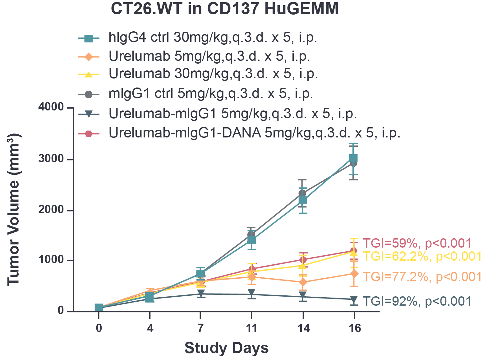
Fig 1. The tumor growth inhibition of urelumab and chimeric urelumab on murine colon tumor CT26.WT in CD137 HuGEMM model. Tumor growth inhibition (TGI) was calculated as: TGI% = (1-Ti/Vi)*100; Ti as the mean tumor volume of the treatment group on the measurement day; Vi as the mean tumor volume of control group at the measurement day.
Fig 2. Measurement of liver toxicity based on ALT (alanine aminotransferase) and AST (aspartate aminotransferase) levels in CD137 HuGEMM animals. Fasting serum level of ALT (alanine aminotransferase) and AST (aspartate aminotransferase) were measured 2 and 7 days after final dose. One way ANOVA *, **, and *** refer to p<0.05, p<0.01, and p<0.001, respectively.
Fig 3. Histological assessment of liver inflammation following urelumab and chimeric urelumab treatment in CD137 HuGEMM in vivo model. H&E staining of liver tissue. Liver inflammation was evaluated on a scale of 0-3: 0 = none; 1 = mild; 2 = moderate; 3 = severe.
Fig 4. Infiltration of CD45+ immune cells in the liver following urelumab and chimeric urelumab treatment in CD137 HuGEMM in vivo model, as indicated by IHC staining - CD45+ cell density were measured by HALO v3.0.311.363.
Tell us about your needs
© 2024 Crown Bioscience. All Rights Reserved.
© 2024 Crown Bioscience. All Rights Reserved. Privacy Policy
2024-04-23
2021-10-23
site_page