<img height="1" width="1" src="https://www.facebook.com/tr?id=1582471781774081&amp;ev=PageView &amp;noscript=1">

Digital Pathology

A morphology, imaging & pathology-based biomarker analysis platform

Rapidly understand your drug’s mechanism and effect through automated immunohistochemistry and immunofluorescence staining and digital analysis. Comprehensive tissue histology characterization from our stringently validated histology platform allows you to save time without compromising accuracy with our well-validated digital pathology platform to ensure high quality analysis of your preclinical and non-CLIA regulated samples:

Digital Pathology Platform


  • Accelerate your research with our selection of over 400 IHC validated markers and multiplex panels for fast and accurate analysis.
  • Streamline your workflow with our integrated digital pathology platform, offering:

    • Automated staining with Leica BOND RX and Roche Ventana BenchMark ULTRA.
    • High-throughput imaging via Akoya Biosciences’ PhenoImager® HT and 3DHISTECH Pannoramic Scan.
    • Detailed tissue analysis with the HALO® image analysis platform, featuring real-time data sharing and remote review capabilities with HALO Link.
  • Count on our thorough validation and QC process for consistent and reliable data.
  • Draw on our immunology and immuno-oncology expertise using translational tumor models based biomarker analysis to gain insights into the tumor microenvironment and the efficacy of cancer treatments.

Download validated markers and panels list

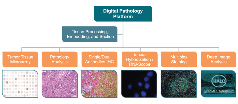
DISCOVER OUR TUMOR TISSUE MICROARRAY COLLECTION


examples_digital_pathology

Akoya Biosciences’ PhenoImager® HT

Advanced Multispectral Imaging (MSI) Technology

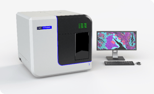
PhenoImager® HT (formerly Vectra® Polaris™) is the fastest and most highly cited whole-slide, single-cell resolution imaging platform for spatial phenotyping and the development of spatial signatures. Featuring Akoya’s patented Multispectral Imaging (MSI) and spectral unmixing technologies, this platform can be easily integrated into high-throughput workflows to accommodate projects regardless of your scale.


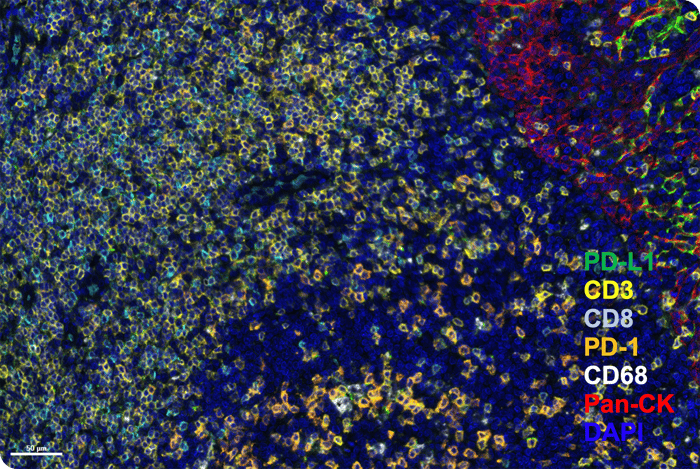
Figure. PD-L1-Immune Panel--Human Tonsil

How PhenoImager HT Works:

  • Multispectral Unmixing: It discerns specific biomarkers from autofluorescence and overlapping signals.
  • High-Throughput Workflow: From custom assay development to multiplex imaging, brightfield and fluorescence imaging up to 9 colors.
  • Automation: The PhenoImager HT offers a fully enclosed system with touchless slide handling for a streamlined workflow.

Benefits at a Glance:

  • Clarity and Precision: The technology unmasks up to 8 biomarkers simultaneously, providing clear, quantifiable insights without autofluorescence interference.
  • Speed and Scale: Conduct comprehensive 6-plex whole slide scans in under 20 minutes with an 80 slide capacity, bolstering high-throughput operations.
  • Versatile Imaging: Whether it's immunofluorescent or immunohistochemical stains, the PhenoImager HT delivers detailed images across a range of staining techniques.

Request a Quote