- Services
- Therapeutic Areas
- Model Systems
- In Vitro
- In Vivo
- Technologies
- Service Type
- About Us
- Our Science
- Start Your Study Now
High-frequency (HF) ultrasound guided injection and monitoring of orthotopic murine models is an alternative to surgical methods that provides more precise implantation, in-life tumor tracking, and improved animal welfare.
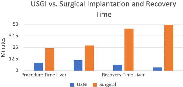
HF ultrasound guided implantation offers higher throughput when compared to surgical implantation due to the shorter procedure time, need for fewer technicians, and faster animal recovery time. The end result is a shortened study duration when compared to surgical implantation.
The use of HF ultrasound imaging of orthotopic models to track tumor progression has several advantages over traditional imaging modalities used to track bioluminescently-tagged tumors. This method of tracking tumors in vivo is non-invasive, does not require the use of tagged cell lines, and allows for 2D and 3D imaging for tumor tracking and precise measurement of tumor volume and location.
HF ultrasound in orthotopic mouse models is a promising tool that provides accurate, reproducible and translational insights for today's therapeutic development.
| Features | Benefits | Case Study |
Crown Bioscience has selected the Vevo 3100 HF ultrasound (Fuji Film Visual Sonics). This is a micro-ultrasound technology that offers high frequency to produce higher resolution. The frequency range of 12 – 71 MHz and 30-micron resolution are well-suited for small animal studies. The high resolution of the Vevo 3100 HF ultrasound allows for precise and less invasive orthotopic tumor implantation using ultrasound guidance, as well as accurate tumor detection and sizing in 2D and 3D imaging. Crown Bioscience has established animal protocol licensing to expand to other orthotopic organs/regions of interest.
Orthotopic implantation of cell lines can be time consuming, technically challenging and inefficient due to the laboratory and surgical procedures required to establish these tumor models. The utilization of HF ultrasound for orthotopic tumor implantation and monitoring overcomes many of these challenges and offers many benefits;
Comparison of HF ultrasound to surgical implantation and traditional monitoring
HF ultrasound guided implantation offers higher throughput when compared to surgical implantation due to the shorter procedure time, need for fewer technicians, and faster animal recovery time. The end result is a shortened study duration when compared to surgical implantation. Results of a comparison study of ultrasound guided implantation (USGI) versus surgical implantation conducted with liver and pancreas tumors is shown in the figure below.
Tell us how we can help!

Factsheet
High-frequency UltrasoundGuided Injection andMonitoring of OrthotopicMurine Models

White Paper
Imaging Orthotopic and Metastatic Preclinical Tumor Models

Webinar
Adding Value to Drug Development Through In Vivo Model Imaging

![]()
![]()
