- Our Services
- Platforms
- Target Solutions
- Technologies
- Service Types
- Our Science
- About Us
- Contact us
Chris Payne, Yasmin Amer, Cathrine Beatty, Ganisha Hutchinson, Mingfa Zhang, Lucy Harris, Amanda Miles, Ruth Storer, Yinfei Yin 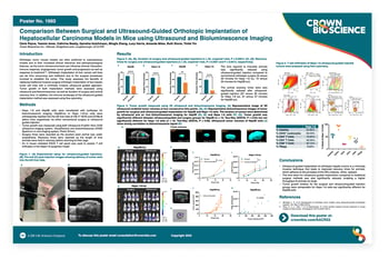 Orthotopic tumor mouse models are often preferred to subcutaneous models due to their increased clinical relevance. However, orthotopic implantation of some cell lines can be time consuming and inefficient due to the surgical procedures involved to establish the tumor.
This study assessed the benefits of replacing traditional invasive surgical orthotopic implantation of hepatic tumor cell lines with a minimally invasive ultrasound guided approach. Tumor growth of both implantation methods were assessed using ultrasound and bioluminescence, as well as duration of surgery and animal recovery time. In addition, the immune landscape of both implantation methods was assessed using flow cytometry.
![]()
![]()
