
Murine Tumor Homograft Models TMA001
H&E and IHC staining
Source: Tumor Homograft Models primary tumor homografts
Catalog #: TMA-MP-MX-001
This Tumor Homograft Models tumor microarray is derived from primary tumor homografts. Each tumor sample is formalin fixed, paraffin embedded, and spotted in triplicate on the TMA.
Have a Question?
Tumor Microarray Profile
| Catalog # | TMA-MP-MX-001 |
| Model Type | Tumor Homograft Models primary tumor homografts |
| Host Species | Mouse |
| # Models On TMA | 12 |
| # Cores/Model | 4 |
| # Cores On TMA | 48 |
| Core Size | 2 mm diameter |
| Fixation Method | Formalin fixed, paraffin embedded |
| Applications | H&E and IHC staining |
| Storage Conditions | 4°C Stable for 12 months from date of purchase when stored as recommended |
| Slide Preparation | Slides are sealed with wax; heat slide at 60° for 1 hour before de-waxing |
| QC | Each TMA slide has >90% core occupancy. H&E and Ki67 IHC staining have been performed on a representative slide of each lot of TMA product. |
Tumor Microarray Map
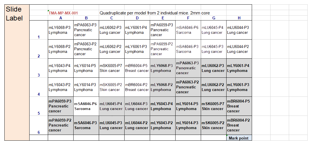
Map of tumor microarray slide
Please follow standard IHC staining procedure.
×
