Explore the Power of High Content Imaging
High content imaging (HCI) and analysis of 3D in vitro models, such as organoids and spheroid cultures, provide invaluable information that gives researchers greater insights into complex disease pathophysiology for more effective assessment of novel compounds and new therapeutic modalities.
Crown Bioscience offers a suite of powerful 3D in vitro imaging-based assays that recapitulate and quantify complex human biology in a robust and high-throughput imaging platform.
These HCI assays are ideal for testing compounds for oncology, immuno-oncology, inflammation, and cystopathy indications. Coupled with our growing organoid biobank of PDX-derived organoid (PDXO) tumor models and patient-derived organoids (PDO), our 3D in vitro high content imaging services enable you to discover the full therapeutic potential of your compounds and new modalities.
Key advantages include:
- High content 3D imaging for phenotypic evaluation of response to treatment in a customizable and scalable 384-well format
- 3D image analysis with highly specialized high content imaging analysis software designed to measure >500 phenotypic changes induced by your compound of interest
- Reliable, reproducible data provides clinically relevant information on compound effects to support important decisions throughout your drug discovery and development workflow— and test multiple drug combination strategies simultaneously
- Physiologically relevant 3D in vitro environments. Services are available for Crown Bioscience’s extensive biobank of well-characterized PDXO and PDO models with patient data. 3D spheroid models from CDX and PDX material are also available
Learn more about our HCI Services for high-throughput compound screening, immuno-oncology, and cystopathic diseases.


Detect therapeutic responses to compounds, antibodies, growth factors, and combinations, such as activity, toxicity, synergy, as well as mode of action and off-target effects. Models are seeded in a hydrogel to allow 3D outgrowth. Drug treatment protocols may be conducted in proliferating or in established tumoroids and spheroids.
Key advantages include:
- Customizable and scalable high-throughput screening (HTS) platform (384-well plate format) in combination with high content imaging readouts
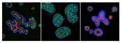
- Measurement of > 500 features such as number, shape & size of nuclei, organoid & lumen formation, or infiltration of immune cells
- Quantitative analysis of growth, morphology, proliferation, cell cycle arrest, cell death, apoptosis, and toxicity
- Functional testing of antibodies, ADCs, growth factors, cytokines, immune cell interactions, and small molecules.
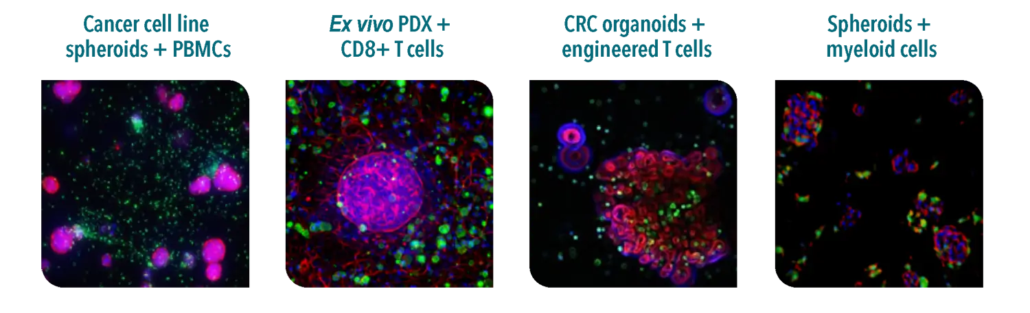
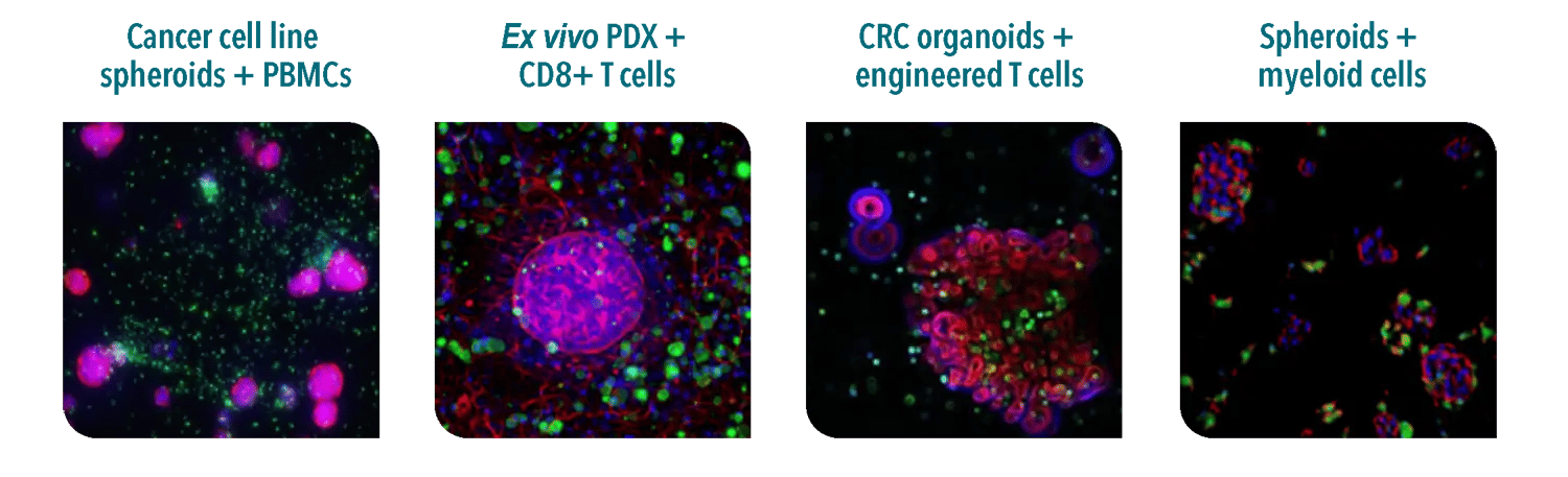
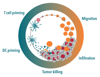

Test the ability of compounds to potentiate migration and infiltration of T cells into tumors, enhance cytotoxic activity against proliferating cancer cells, repolarize immunosuppressive myeloid cells, and study the interactions with activated immune cells in immuno-oncology organoid cultures.
Key advantages include:
- Automated analysis and robust quantification of activity of immune cell populations
- Functional readouts: immune cell priming, active migration, infiltration into tumoroids, tumor cell killing, and myeloid polarization
- Physiologically relevant 3D microenvironment
- Visualization of immune cell interaction with the tumor
- HLA-matched cell types available