
Our standard flow cytometry panels have been validated in multiple oncology and inflammation in vivo models to provide basic to comprehensive analysis of lymphoid and myeloid cell populations. Models analyzed include syngeneics, tumor homograft models, human target expressing (Humanized genetically modified mouse models / Humanized Target Tumor Cells), humanized, GvHD, and peritonitis models.
Advanced High-Parameter Analysis: Spectral Flow Cytometry
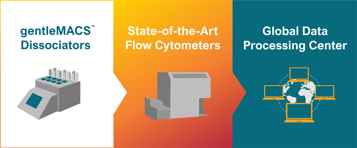
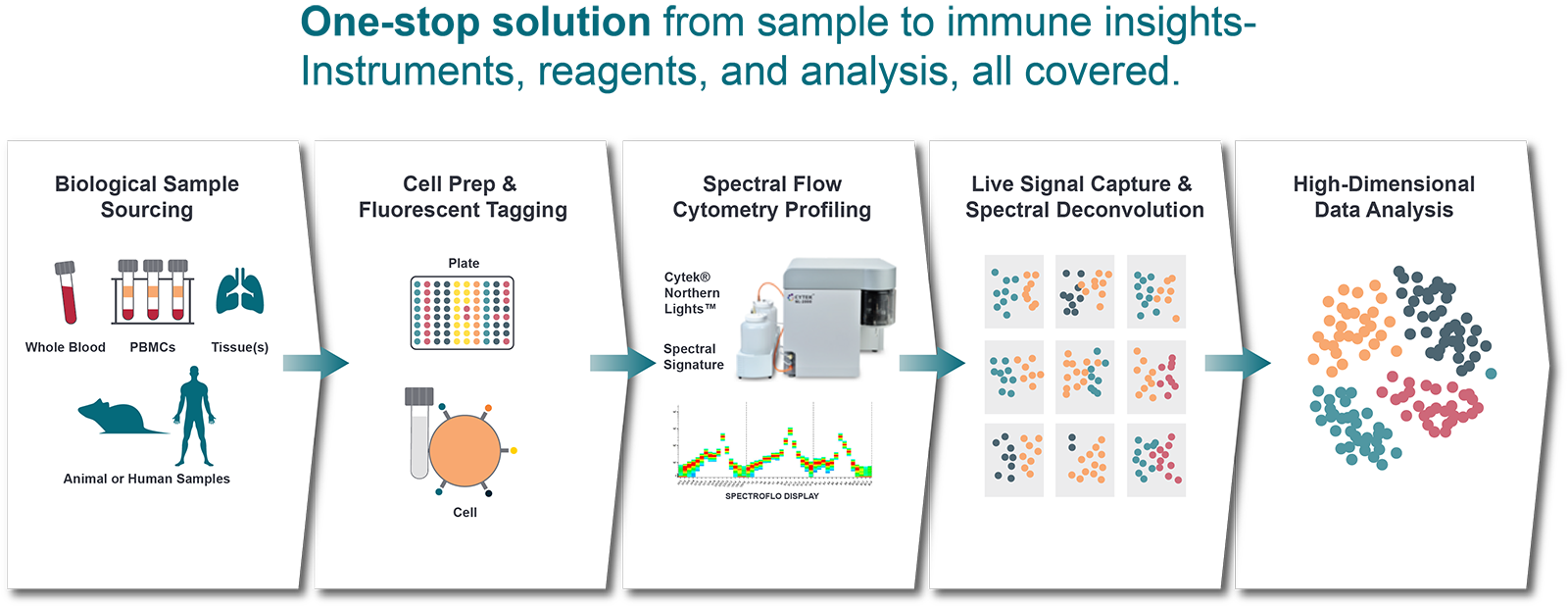
Unlock unprecedented depth in cellular analysis with Crown Bioscience's state-of-the-art Spectral Flow Cytometry services. Moving beyond conventional flow cytometry, spectral flow utilizes the entire emission spectrum of fluorophores, rather than relying on discrete bandpass filters. This revolutionary approach allows for the simultaneous detection and analysis of a significantly higher number of markers (typically 20-40+ parameters), providing a truly comprehensive view of complex biological systems.
Key Advantages for Your Research:
- Unrivaled Parameter Capacity: Characterize intricate cellular populations and rare cell types with unparalleled resolution, essential for dissecting heterogeneous samples like tumor biopsies or complex immune repertoires.
- Enhanced Data Quality: Minimize spectral overlap (spillover) and improve signal resolution, leading to cleaner, more accurate data for even dimly expressed markers.
- Fluorophore Flexibility: Utilize a broader range of fluorophores, including those with highly overlapping emission spectra, optimizing panel design and expanding experimental possibilities.
- Maximized Sample Insight: Extract more information from precious clinical or research samples, reducing the need for multiple assays and preserving valuable material.
- Streamlined Analysis: Advanced spectral unmixing algorithms simplify data interpretation, making high-dimensional data more accessible and actionable.
Applications for Cancer Researchers:
Leverage spectral flow cytometry to:
- Deeply Phenotype the Tumor Microenvironment (TME): Precisely characterize immune cell subsets (T-cells, B-cells, NK cells, myeloid cells, etc.) and their activation states within tumors, peripheral blood, and lymph nodes.
- Accelerate Biomarker Discovery & Validation: Identify novel cell-surface and intracellular biomarkers for patient stratification, prediction of therapy response, and disease progression, by correlating complex phenotypic profiles with clinical outcomes.
- Elucidate Drug Mechanisms of Action (MOA): Gain granular insights into how novel therapeutics impact target cells, immune populations, and resistance mechanisms.
- Monitor Immunotherapy Responses: Track subtle changes in immune cell composition and function over time in patients receiving immunotherapies, providing crucial insights into treatment efficacy and potential adverse events.
- Analyze Rare Cell Populations: Confidently detect and characterize rare tumor-infiltrating lymphocytes (TILs) or circulating tumor cells (CTCs) with high specificity and sensitivity.
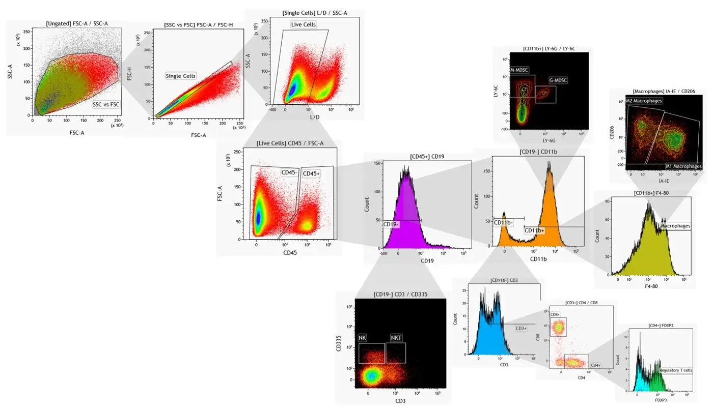
Customized panels are also available, which undergo thorough validation prior to running your samples. See our standard panels and gating strategy below.
| Markers | Immune Cell Population |
|---|---|
| CD45+ | Total leukocytes |
| CD45+ CD11b- CD3+ | Total T cells |
| CD45+ CD11b- CD3+ CD4+ CD8+ | CD4+ T helper cells |
| CD45+ CD11b- CD3+ CD4+ CD8+ | CD8+ cytotoxic T cells |
| CD45+ CD11b- CD3+ CD4+ CD8+ Foxp3+ | Regulatory T cells |
| CD45+ CD3- CD11b+ Ly-6C+ Ly-6G- | M-MDSC |
| CD45+ CD3- CD11b+ Ly-6C- Ly-6G+ | G-MDSC |
| CD45+ CD11b+ Ly-6C- Ly-6G- F4/80+ | Macrophages |
| CD11b+ F4/80+ Gr-1+ | MDSC |
| CD45+ CD11b+ Ly-6C- Ly-6G- F4/80+ IA-IEhigh CD206low/- | M1 macrophages |
| CD45+ CD11b+ Ly-6C- Ly-6G- F4/80+ IA-IElow/- CD206+ | M2 macrophages |
| CD45+ F4/80- Ly-6C- Ly-6G- CD3- CD335+ | NK cells |
| CD45+ F4/80- Ly-6C- Ly-6G- CD3dim CD335+ | NKT cells |
| CD19 | B cells |
| PD-1 / PD-L1 | Checkpoint inhibitors |
| Live / Dead (fixable) | Live / Dead |
Basic panel comprises of 10 colors
Bold markers are included in the comprehensive 16 color panel
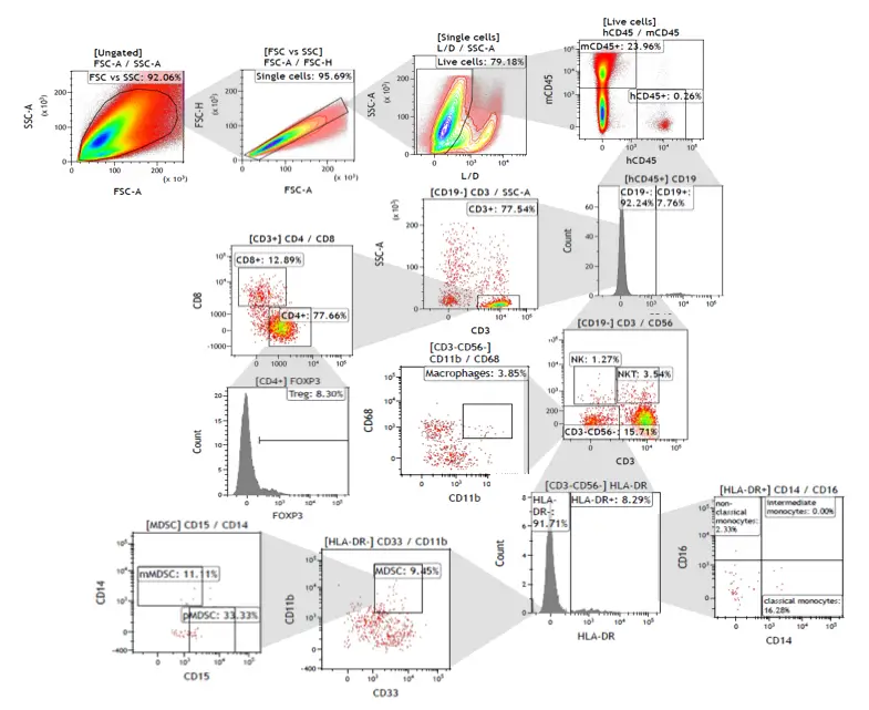
| Markers | Immune Cell Populations |
|---|---|
| mCD45-/hCD45+ | Total leukocytes |
| mCD45-/hCD45+/CD3+ | Total T cells |
| mCD45-/hCD45+/CD3+/CD4+ | CD4+ T helper cells |
| mCD45-/hCD45+/CD3+/CD8+ | CD8+ cytotoxic T cells |
| mCD45-/hCD45+/CD3+/CD4+/Foxp3+ | Regulatory T cells |
| mCD45-/hCD45+/CD19-/CD3-/CD56+ | NK |
| mCD45-/hCD45+/CD19-/CD3+/CD56+ | NKT |
| mCD45-/hCD45+/CD19-/CD3-/CD56-/CD11b+/CD68+ | Macrophages |
| mCD45-/hCD45+/CD19-/CD3-/CD56-/HLA-DR+/CD14+CD16- | Classical monocytes |
| mCD45-/hCD45+/CD19-/CD3-/CD56-/HLA-DR+/CD14+CD16- | Intermediate monocytes |
| mCD45-/hCD45+/CD19-/CD3-/CD56-/HLA-DR+/CD14-CD16- | Non-classical monocytes |
| mCD45-/hCD45+/CD19-/CD3-/CD56-/HLA-DR-/CD33+CD11b+ | MDSC |
| mCD45-/hCD45+/CD19-/CD3-/CD56-/HLA-DR-/CD33+CD11b+/CD14+/CD15- | M-MDSC |
| mCD45-/hCD45+/CD19-/CD3-/CD56-/HLA-DR-/CD33+CD11b+/CD14-/CD15+ | P-MDSC |
| Live / Dead (fixable) | Live / Dead |
Characterize the biological responses of immune cells through flow cytometry based functional assays including receptor occupancy, apoptosis, and proliferation assays.
