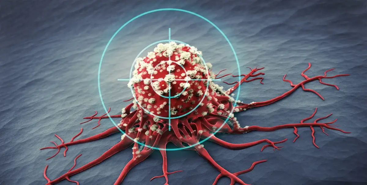
Metastases form when tumor cells shed from their primary site and circulate in the bloodstream before leaving the vasculature to colonize distant viable organs. These circulating tumor cells constitute seeds for subsequent growth of secondary tumors that are responsible for the vast majority of cancer related deaths.
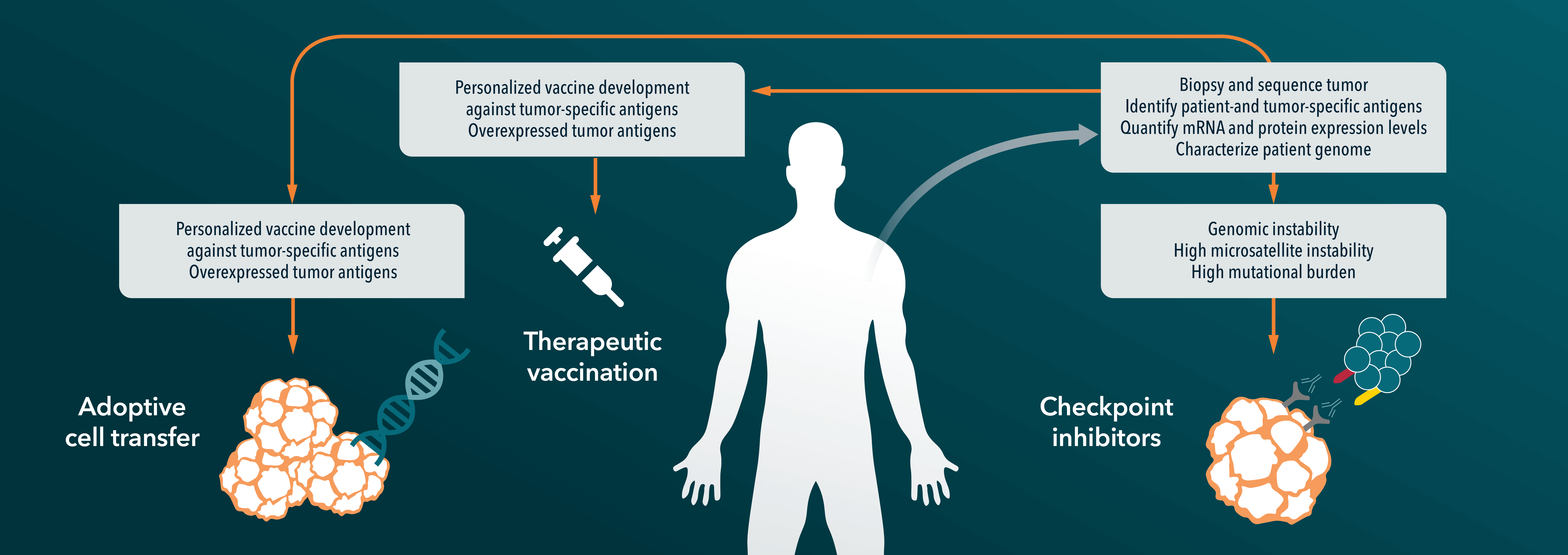
Circulating tumor cells (CTCs) were observed for the first time in 1869 by Thomas Ashworth who understood that they originated from the primary tumors and represented the “origin of multiple tumors existing in the same person”. The importance of CTCs in modern medicine is underlined by the blossoming of liquid biopsy techniques during which a patient’s blood is analyzed in search of viable circulating cells, with an intact nucleus, that show the same characteristics (biomarkers) as the patient’s tumor. Blood tests are easier and safer to perform than conventional biopsies, and multiple samples can be taken over time, facilitating appropriate modification to a patient's therapy regimen, potentially improving their prognosis and quality of life. Liquid biopsies have contributed to the understanding that tumors are heterogeneous entities composed by cells carrying different mutations.
During the recent American society of clinical oncology (ASCO) annual meeting, the participating oncologists discussed new applications, clinical utility data, and efforts in optimizing readouts or automation of liquid biopsies for CTCs and circulating tumor DNA (ctDNA). An abstract submitted to the meeting and released online described a study by Biocept, a diagnostics developing company specialized in liquid biopsies, in which researchers found in one patient’s blood sample an EGFR mutation that was not present in the corresponding lung tumor. Puzzled by this difference, they performed a second biopsy of the primary tumor and confirmed the presence of the same mutation in another part of the tumor, allowing the patient recruitment within the relevant clinical trial.
In a recent report published in Nature Medicine researchers from the University of Turin genotyped ctDNA from colorectal cancer patients subjected to EGFR inhibitors (EGFRi)-based therapy. By this means they were able to follow the evolution of the primary tumor as patients underwent therapy and to identify the genetic alterations occurring while drug resistance emerge. The results also explain at a molecular level why patients seem to benefit from “treatment holidays”, periods in which EGFRi-based therapy is temporarily interrupted.
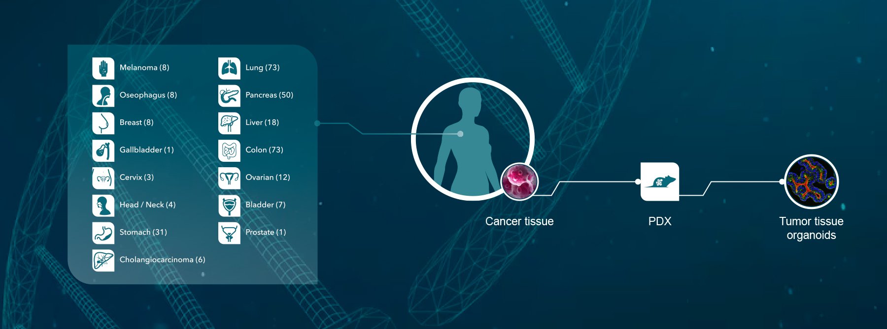
At Crown Bioscience we strive to be at the forefront of cancer research. To provide a robust platform for preclinical evaluation of therapeutic efficacy of new and existing anticancer drugs, appropriate in vivo models are required that recapitulate the results observed in the clinic. To tackle the role of CTCs in the development of drug resistance in cancer patients we have established a human gastric carcinoma PDX model that shows high potential of lung metastasis. Our models displays large and small size CTCs circulating as single cells or small aggregates – called tumor microemboli (CTM) – and their abundance correlates with the number of lung metastatic lesions. CTCs respond differently to chemotherapy depending on their genetic composition with multiploid cells displaying drug insensitivity. Our data represent the first attempt to combine the morphological examination of the metastatic lesion and the genetic karyotyping of the CTCs with in vivo data on the response of the animal to chemotherapy, proving that metastatic PDX model (mPDX) displaying CTCs are of particular significance for translational studies.
Crown Bioscience can be contacted at busdev@crownbio.com for further information on our platforms for CTCs research or on any other of our products.


